Showing posts with label Anemia. Show all posts

Wednesday, October 15, 2008

Normocytic Anemia


Normocytic, normochromic anemia is the largest, most frequently encountered anemia. If acute blood loss can be ruled out, the diagnosis remains to be intrinsic or extrinsic. Hereditary spherocytosis, drug induced anemia, and anemia secondary to other malignancies are a few of the anemias of this classification.

Normocytic anemia may include hemolytic anemia. The peripheral blood smear and the history often suggest possible causes for the anemia.


Normocytic Normochromic - Hemolytic Anemia


Sickle Cell Anemia


Microcytic Anemia

Microcytic anemia is based on the protocol for patients who have an automated MCV of less than 75 fl. The three most common causes of microcytic anemias are iron deficiency, thalassemia minor, and anemia of chronic disease. Cases of thalassemia have elevated red counts and lower RDW's than would be expected for the MCV and the degree of anemia. Iron deficiencies are almost always associated with a high RDW. Some anemias of chronic disease may be normocytic and others such as renal disease are microcytic. Anemias of chronic disease do not have decreased iron stores.


Microcytic Anemia - Hypochromic, Target Cells, Microcyte


Macrocytic Anemia


Macrocytic anemias are less commonly encountered than normocytic or microcytic anemias. These anemias may be caused by marrow failure such as aplastic anemia and myelodysplasis, or caused by deficiencies of vitamin B12 or folic acid; or caused by autoimmune hemolysis or cold agglutinins. A mild degree of macrocytosis with a normal RDW is commonly seen as a result of alcohol abuse. The MCV is greater than 100 fl. in macrocytic anemia.


Peripheral Blood


Bone Marrow


The macrocytic anemias may be further subdivided based upon the degree to which the MCV is raised and the presence of megaloblastic production in the bone marrow.

Slight increase in MCV:

MCV > 100

  • due to the presence of retics
  • in some instances of aplastic anemia
  • myxedema

In all cases the red cell precursors in the marrow are normal in morphology.


Moderate increase in the MCV:

MCV > 105

  • liver disease


Marked increase in the MCV:

MCV > 110 fl.

  • megaloblastic due to the lack of vitamin B-12 or folic acid.


Thursday, September 18, 2008

Classification of Anemia


INTRODUCTION

Anemia is a disorder that results in a decrease in the ability of the blood to carry oxygen. Anemia is itself not a diagnosis but merely a sign of underlying disease. The initial classification of anemia is best accomplished by examination of the data from a hematology analyzer and by an examination of the peripheral blood smear. The physician most commonly classifies anemias initially by the instrument's red cell indices, especially the mean corpuscular volume (MCV). On newer counters, the red cell distribution width (RDW) or red cell morphology index (RCMI) is another useful measurement. The anemia may be microcytic, normocytic, or macrocytic.


CLASSIFICATION of ANEMIA


Anemia is usually classified according to:

  • Etiology
  • Pathophysiology
  • Morphology

1. ETIOLOGIC

This is classification by cause. An anemia may be due to blood loss which may be due to many causes, eg; excessive vaginal bleeding due to functional menorrhagia, malignancy or endometriosis.

2. PATHOPHYSIOLOGIC

This classification is based on the actual red cell defect like decreased red cell production or increased red cell destruction. In other words the anemia can be classified as:

  • Nonregenerative
  • Regenerative

In many cases either defect are operative or there is uncertainty as to the exact defect which is operative.

3. MORPHOLOGIC

This is a classification based on cell size and color. This classification is usually used in the laboratory as we actually see the cells. It is not entirely satisfactory as an anemia due to chronic bleeding may be normocytic at one point, microcytic later and microcytic hypochromic even later.In fact the most often used classification system is a combination of the pathophysiologic


CONCLUSION


All in all, these classifications will aid in treatment of the disease. It is important to know all these classification so that the diagnosis done will be more accurate and precise. Among the first step of diagnosis is screening. Screening is usually done with the CBC or "complete blood count". The exact procedure in a CBC depends upon the instrumentation in the laboratory. Most laboratories now use automated, multiparameter instruments which will provide results for the following parameters:


hemoglobin

hematocrit

red cell count

MCV

MCH

MCHC

RDW

white cell count

platelet count

MPV

automated differential

histograms and scattergrams


As a person who’s going to be working in the lab, assessing the sample, one should also know how to observe a stained blood film to differentiate one anemia from another. A stained blood film should be examined whenever any of the above parameters are abnormal. Examination of the blood film can identify a large range of erythrocyte, leukocyte and thrombocyte changes. The presence of red cell inclusions indicates abnormal erythropoiesis or an increased rate of red cell destruction while poikilocytes indicate a severe red cell abnormality. In some instances poikilocytes can suggest a specific diagnosis. Changes in leukocytes and platelets can provide a clue as to the etiology of an anemia. Some of these changes will be detected by current automated instrumentation. Once a tentative diagnosis is made special tests can be performed to confirm the diagnosis. These include iron studies, vitamin studies, hemoglobin studies, enzyme levels, bone marrow aspirate or biopsy, red cell survival studies etc.

Plus, one should make a thorough diagnosis so that errors can be avoided. Some of the errors are; errors in reporting or recording of results, inadequate study of the blood film, failure to assess indices, failure to do retic count, failure to note rouleux or blue background on film.

Wednesday, September 17, 2008

Sideroblastic Anemias (SA)


Sideroblastic anemias (SA) are described as a heterogeneous group of anemias. There are three general types of SA, acquired, idiopathic, and hereditary. Regardless of the type of pathogenic mechanism, all types of sideroblastic anemias are characterized by:

  • Increases in total body iron
  • The presence of ringed sideroblasts in bone marrow.
  • Presence of microcytic, hypochromic RBC’s.
  • Decreased MCH and MCHC values.
  • Decreased Retic count.

Type 1: Hereditary Sideroblastic Anemia (SA)


Type 1: Hereditary Sideroblastic Anemia (SA)

A sex-linked, recessive disorder primarily affecting males and is the more severe form. This form is not common and tends to be progressive, requiring transfusion support. This is a recessive autosomal-linked SA. This usually appears after the age of seven, but can manifest in infancy. It is very rare but this form has been reported as manifesting symptomatically around the age of sixty.

A few of these patients may demonstrate hepatomegaly.

Clinical laboratory findings include:

  • WBC count is usually normal, but may be decreased
  • Platelet count is variable.
  • Reticulocyte Production Index is usually <2.0.
  • A moderate to severe anemia is present with a dimorphic RBC’s in the stained blood film.
  • Both normochromic, normocytic and hypochromic, microcytic RBC’s are observed.
  • A few patients may demonstrate mild macrocytosis.
  • If the blood smear appears to have an equal distribution of microcytes and normocytes, the indices will usually be within the normal range.
  • Target cells are prominent.
  • Anisocytosis and poikilocytosis may be present.
  • Both Pappenheimer bodies and basophilic stippling may be observed. These must be differentiated from each other.

Type 2: Idiopathic Sideroblastic Anemia (IASA)

Type 2: Idiopathic Sideroblastic Anemia (IASA)

  • This is a more common form of S.A., occurring more frequently than Type One. It usually occurs during adulthood.
  • The cause is not known, but has a predilection for patients >50 years old. It is often found when the patient visits the physician for some other supposed condition. This is a primary anemia and is also called “Refractory Anemia with Ringed Sideroblasts (RARS) because of the presence of ringed sideroblasts.
  • It is considered to be a form of myelodysplastic syndrome.
  • Two or more blood cell lines may be affected which may represent a refractory anemia with a form of a chronic-like leukemia. This occurs in a few patients and has a poor prognosis. Up to 10% of the patients with IASA may develop acute leukemia.
  • The peripheral blood may present with a dimorphic RBC population in which anisocytosis, poikilocytosis, hypochromia, and basophilic stippling may be observed.
  • One indicator of this variant is the bone marrow study that presents with >15% of the NRBC’s in the form ringed sideroblasts (and may make up 90%).
  • A number of patients do not require medical intervention because the disease is non-progressive and benign. If the bone marrow of these patients presents with >30% ringed sideroblasts with abnormal granulopoiesis and megakaryocytopoiesis, the prognosis becomes poor.
  • IRSA appears similar to erythroleukemia. To differentiate, the IASA rubriblast is periodic acid-Schiff negative whereas for erythroleukemia it is positive.
  • Clinical laboratory findings are somewhat similar to other types of S.A.

Type 3: Acquired Reversible Sideroblastic Anemia(ARSA)


A secondary form of anemia, where the cause is resultant of another problem.

Drugs/medications are the more common causes of this anemia:

  • Isoniazid, an anti-tubercular drug, inhibits reactions requiring pyridoxal-5'-phosphate as a coenzyme.
  • Chloramphenicol: anti-bacterial, blocks mitochondrial protein and heme synthesis.
  • Cytotoxic drugs and poisons probably work similar to isoniazid and chloramphenicol.
  • Copper deficiency has been reported to contribute to the development of this anemia.

Infections and leukemias in context with the medications used as therapeutic measures.

If the causative agent is removed, then the anemia is reversed.

Ethanol ingestion (alcoholism): Blocks heme synthesis.This form of anemia is seen in 30% of alcoholics admitted to the hospital. Lead poisoning induces several effects, one of which is sideroblastic anemia.

  • Lead consumption, regardless of the route, interferes with iron storage in the mitochondria inducing classical S.A.
  • Lead also damages a minimum of six enzyme required for the synthesis of hemoglobin. The life span of the RBC is shortened. The enzyme “δ-aminolevulinic dehydrase” is most sensitive to lead of all the enzymes. δ-aminolevulinic acid is elevated in lead poisoning, making it a good indicator for lead toxicity.

Clinical laboratory findings that indicate lead poisoning are:

  • Basophilic stippling
  • Reticulocytosis
  • Leukocytosis with increased eosinophils
  • Presence of reactive lymphocytes
  • Increased Hemoglobin F

Iron studies of sideroblastic anemia tend to demonstrate the following:

  • High plasma and tissue iron content.
  • Free erythrocyte protoporphyrin is increased. This is due a defect in incorporating Fe++ into the protoporphyrin IX molecule to form heme.
  • The TIBC will be low or normal. The high plasma iron concentration keeps the iron bearing proteins saturated.
  • Serum ferritin is increased.
  • Percent iron saturation of ferritin is increased.

When examining the bone marrow of a sideroblastic anemic patient you may see:Presence of erythroid hyperplasia. Megaloblastosis MAY BE present. The degree of megaloblastosis is dependent upon the severity of the anemia. Megaloblastosis is usually associated with a deficiency in vitamin B12 and/or folic acid.

  • If megaloblastosis is present, then the giant erythrocyte precursors will give rise to the following maturation sequence:megaloblastic rubriblast, megaloblastic prorubricyte,megaloblastic rubricyte,megaloblastic metarubricyte,macrocytic
  • polychromatophilic cell,macrocyte.
  • Aggregates of iron granules may be observed in the cytoplasm.
  • The normal RBC series (rubriblast, prorubricyte, rubricyte, metarubricyte) may present a cytoplasm that appears to be poorly hemoglobinized. The cytoplasm may also appear scanty and frayed.
  • Macrophages may be observed and may contain increased amounts of storage iron.
  • Presence of sideroblasts. This is the distinguishing feature of this type of anemia. A normal rubriblast may contain up to five small granules of iron. If more than six granules of iron are observed then this may be considered a pathological condition. The iron granules can be demonstrated using Perls’ Prussian blue iron stain.

Early normal immature erythrocytes (this range as been cited from a low 20% to a high of 80%) will contain an occasional free siderotic granule located in the cytoplasm, but not in any organelle. Some textbooks set the range from 30 to 50%, which may be more realistic.

In sideroblastic anemia, look for 10% to 40% of the RBC precursors to contain increased siderotic granules around the nucleus. If the siderotic cells are predominately early RBC precursors, then this is probably a hereditary form of sideroblastic anemia. If the affected cells include the late precursor cells, then this would be an indicator of an acquired form of sideroblastic anemia. Note that target cells are usually not observed in stained blood films.

Iron-Deficiency Anemia (IDA)

Anisocytosis with hypochromia and microcytes (IDA)

Iron-deficiency anemia (IDA) is the most commonly encountered anemia and may be due to [1] impaired iron intake, [2] pregnancy, [3] intravascular hemolysis, [4] hemorrhage, or [5] lactation. IDA most often affects women in their reproductive years and growing children. Each mL of packed RBC’s contain about 1.0 mg of iron. The average adult contains about 3.5 to 5.0 grams of iron. They will ingest about 15 to 20 mg of iron daily, excreting most of it. The body normally absorbs about 1 mg of iron (which is equal to the daily loss).


The iron taken into the body is in the ferric state (Fe+3) in the stomach, it is changed to the ferrous state (Fe+2). Ferrous iron is absorbed by the small intestine and in the intestinal capillary system, iron is bound to transferrin to form a protein-iron complex. This complex is carried to the bone marrow (and other cells requiring iron) and will bind to the cell. The complex is absorbed into the cell, the iron released, and the transferrin moves back into the blood stream. Inside the cell, the iron is bound to the protein apoferritin to form ferritin. The ferritin combines to form aggregates which forms brown pigment granules called hemosiderin. IDA is a hypoproliferative, microcytic, hypochromic anemia due to ineffective RBC and/or hemoglobin production.


If IDA is observed in a healthy appearing adult male, the physician should look for a gastrointestinal lesion that may be losing blood. The menstruating woman will lose between 50 and 70 mLs of blood monthly and when the iron is not being replace anemia will result. It is estimated that up to 20% of the women in the US have IDA. IDA produces a hypochromic, microcytic anemia. On the peripheral blood smear, the erythrocytes are hypochromic and microcytic. If IDA is severe, poikilo-cytosis and anisocytosis may be obvious. In the bone marrow, the rubriblasts will be poorly hemoglobinized and demonstrate ragged appearing cytoplasm. The serum iron will be decreases and the total iron binding capacity (TIBC) will be increased. The ferritin level will be less than 20 ng/mL.


IRON TRIVIA


If dietary iron is from meat sources, it is heme bound. Vitamin C is not required for absorption. If this iron is from eggs and vegetables, it is in the ferritin and hemochrome bound form and requires vitamin C for optimum absorption. In the stomach, the gastric fluid and pepsin releases iron which passes into the gastrointestinal tract. Most iron absorption occurs in the duodenum. Some iron absorption will occur in the jejunum and ileum.


In developed countries, adequate ion intake is not a problem. The high risk groups who are most likely to develop IDA are [1] infants, [2] rapidly growing adolescents, [3] pregnant women, and [4] women during their child bearing years (losing from 10 to 45 mg/month). The pregnant female need about 3.4 mg of iron daily (a total of 1000 mg to carry the fetus to term). About 400 mg are needed for the fetal RBC mass. At parturition, approximately 300 mg will be lost and up to 170 mg will be contained in the placenta and umbilical cord.


It has been estimated that a healthy adult male would require about eight years to develop IDA if no more iron were absorbed in his diet. Malabsorption is uncommon unless there is a primary problem as [1] sprue, [2] gastrectomy, or [3] atrophic gastritis. Other causes are [1] regular blood donations and [2] paroxysmal nocturnal hemoglobinuria.


CLINICAL SYMPTOMS


Early stages (stage 1) are generally asymptomatic. As IDA develops into stage two, the depletion of the body’s iron stores occurs, with the patient experiencing hypoxia, characterized by lethargy and asthenia. As stage two progresses, iron deficiency is demonstrated by a decrease in erythropoiesis as iron is no longer being inserted into the hemoglobin molecule. Lab testing will show decreases in serum iron, increased total iron binding capacity (TIBC), and low transferrin saturation.


As the IDA progresses into the stage three level, the mitotic activity of the RBC increases resulting in small erythrocytes (microcytes) and hypochromia. When the hypochromic microcyte is observed in the blood film, there is also anisocytosis and poikilocytosis and IDA if fully expressed. Symptoms that begin to appear in stage two and are fully expressed in stage three are [1] ankle edema, [2] exertional dyspnea, [3] headaches, [4] glossitis, [5] koilonychia, [6] pallor, [7] pica, and [8] tachycardia. In the woman of child bearing age, [1] menorrhagia, [2] irregular cycles, and/or [3] amenorrhea may occur.


CLINICAL LABORATORY FINDINGS


RBC count: Usually normal at the beginning. The count will usually remain within normal limits unless the iron stores are severely depleted.
Hemoglobin: Will undergo greater degrees of reduction. Individual are to be considered anemic if hemoglobin values fall as indicated in g/dL:

  • [1] Children (from 6 months to 5 years) less than 11
  • [2] Children (from 6 years to 14 years) less than 12
  • [3] Adult men. . . . . . . . . . . . . . . . . . . . . . less than 13
  • [4] Adult women. . . . . . . . . . . . . . . . . . . less than 12
  • [5] Pregnant women . . . . . . . . . . . . . . . . less than 11


RBC Indices: Anemia is suspected when the values fall as indicated:

  • [1] MCV . . . . . 75 to 80 fL
  • [2] MCH . . . . . 25 to 27 pG
  • [3] MCHC. . . . less than 32 percent
  • [4] If the MCV is less than 75 fl.
  • [5] If the MCH is less than 25 pG


Retic Count: This test parameter may be normal or decreased in early IDA. As the IDA progresses, the retic count decreases.


The Reticulocyte Production Index is a better indicator of bone marrow responses in anemia.


Fragility: This test will usually be normal. If codocytes (target cells) are present, then one may see a decreased value. The value of this test is in detecting hereditary spherocytosis.


WBC: The count is usually normal as is the differential.


Platelet count: This testing parameter is usually normal.


Serum Iron: decreases in stages as IDA develops.


Normal values (μg/dL) as follows:

  • Newborn . . . . . . . 100 to 250
  • Infant . . . . . . . . . 40 to 100
  • Child . . . . . . . . . 50 to 120
  • Adult male. . . . . . 65 to 170
  • Adult female . . . . 50 to 170


Serum Ferritin: Is an indicator of how much iron is being stored and it will progressively decrease as IDA develops. It is the major iron storage compound and is found in all body cells. It is a protein that is complexed with iron. If iron is absent from this protein, it is then know as apoferritin. This is an important test in differentiating IDA from other types of microcytic normocytic anemias as it will be increased in thalassemia and sideroblastic type anemias. Normal values for men are 15 to 200 μg/L and women are 12 to 150 μg/L.


Free erythrocyte protoporphyrin (FEP): This test provides the same information as the serum ferritin. Protoporphyrin is the compound to which ferrous iron is added to form the heme molecule. In normal hemoglobin production, a little more protoporphyrin is produced than is required. When there is an iron deficiency, protoporphyrin will increase in the RBC. The normal reference is less than 50 μg/dL of RBC.


Generalized test findings as IDA develops.


[1] In the initial stages when patient is asymptomatic:

  • A. Serum ferritin will be decreased
  • B. Bone marrow iron will be decreased


[2] In the second stage, when erythropoiesis is occurring without
iron to insert in the heme portion of the hemoglobin molecule:

  • A. Serum Ferritin continues to be decreased.
  • B. Bone marrow iron continues to be decreased.
  • C. Serum iron is now decreased.
  • D. TIBC is increased.


[3] In the final stage with fully developed IDA:

  • A. Serum Ferritin, bone marrow iron, and serum iron are decreased.
  • B. TIBC is increased.
  • C. Hemoglobin and hematocrit are decreased.
  • D. MCV is decreased.
  • E. RDW is increased.


Bone marrow findings in IDA. NOTE: Bone marrow studies are usually not required to diagnosis IDA.
[1] Mild to moderate erythroid hyperplasia.
[2] Increase in number of cells in the erythroid line.
[3] Look for smaller rubriblasts (normoblasts) with [a] pyknotic nucleus, [b] thin rim of irregular basophilic cytoplasm, [c] the absence of or decreased hemoglobiniz nuclear an[a dding, developed IDA, stainable iron granules are absent.


Megaloblastic Anemia vs Macrocytic Anemia

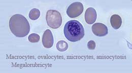
Megaloblastic anemia describes any anemia in which the RBC precursors are of a large size. The earliest recognized immature form is designated as the promegaloblast or megalocytic rubriblast, followed by the megalocytic prorubricyte, megalocytic rubricyte, and megalocytic metarubricyte in the maturation sequence. This cell line occurs because of impaired DNA synthesis due to vitamin B12 and/or folic acid deficiency. This deficiency causes nuclear plasm asynchrony, in which the nuclear maturation events do not occur in sequence nor in the time interval of development they are supposed to. This disruption produces the extra large RBC’s.


The term “megaloblastic” may infer any of the blood cell maturation lines. Megaloblastic anemia is not to be confused with macrocytic anemia. Where megaloblastic anemia is due to faulty DNA synthesis, macrocytic anemia is a secondary problem due to a primary disorder such as [1] liver disease, [2] acute hemorrhage, or [3] severe anemic episode. Macrocytic anemia is characterized by a MCV range of 105 to 115 fL (indicating thin cells or target cells), although the MCV may go from 100 fL to 130 fL and if severe, may be up to 160 fL. The retic count tends to run from 10 to 25% where it is normal for a megaloblastic condition. In megaloblastic anemia the MCV may range from 100 fL to 160 fL with the MCH elevated but the MCHC is usually normal.


Clinical symptoms for the macrocytic and megaloblastic anemia include [1] pallor, [2] mouth soreness, [3] glossitis, [4] lethargy, [5] asthenia, [6] anorexia, [7] weight loss, [8] diarrhea followed by constipation, and [9] headaches. In megaloblastic anemia, the patient may also experience [1] paresthesia (numbness in hands and feet), and [2] changes in proprioception (movement, posture, and equilibrium). If the condition is not corrected, the patient may experience sudden involuntary movements or convulsions and progress to ataxia (defective movements). This is a progressive disease in which the spinal cord may degenerate and/or the patient may become psychotic.


The psychotic stage is known as megaloblastic madness. Megaloblastic anemic disorders are vitamin-dependent of which pernicious anemia describes a condition caused by the absence of intrinsic factor resulting in a B12 deficiency. Other causes may be due to increased requirements for B12 and/or folic acid. Drugs, malabsorption, and dietary deficiencies have been known to contribute to the manifestation of this anemic disorder.

Pernicious Anemia (PA)


Pernicious anemia (a specific megaloblastic anemia), one of the macrocytic anemias, is the consequence of a deficiency of vitamin B12 or its absorption and folic acid. This anemia is caused by impaired DNA synthesis, which leads to the megaloblastic transformation of erythroid precursors. The defective DNA synthesis retards nuclear maturation and causes abnormal mitotic activity which causes the cells to divide too rapidly. The nuclei tend to be large with loosely arranged chromatin material. It is not unusual to observe satellite nuclear pieces in these cells. The cytoplasm maturates at different rates and hemoglobin synthesis is also affected. This abnormal bone marrow environment is characterized by increased cellular destruction and ineffective hematopoiesis.


Cells that are released into peripheral circulation tend to be large and oval in shape. The RBC “picture” may be featured with poikilocytosis and an increased number of dacryocytes. The immature granulocytic cells undergo significant changes. The metamyelocyte tends to be large with a horseshoe shaped nucleus. Neutrophils present with hypersegmentation. The mega-myelocytes tend to be hyposegmented or the nuclear lobes are widely separated. Clinical observations include: [1] increased MCV, [2] macrocytes, [3] anisocytosis, [4] poikilocytosis, [5] hypersegmentation, [6] leukopenia, and [7] thrombocytopenia.


Pernicious anemia (PA), as a rule, only occurs when there is impaired vitamin absorption due to the loss of intrinsic factor, which will combine with the vitamin to form a soluble intrinsic-vitamin B12 complex, where it is absorbed in the terminal portion of the ileum. PA will present with the following symptoms: [1] weakness, [2] pallor, [3] giddiness, [4] anorexia, [5] diarrhea, [6] constipation, [7] glossitis, [8] tachycardia, [9] splenomegaly, and/or [10] hepatomegaly. Causes of defective absorption are; [1] celiac disease, [2] peptic ulcer, [3] stomach cancer, [4] Crohn’s disease, [5] the parasite, Diphyllobothrium latum.


PA occurs frequently in patients with [1] thyroidtoxicosis, [2] Hashimoto’s thyroiditis, [3] rheumatoid arthritis, and [4] gastric carcinoma. Failure to treat this anemia may lead to spinal cord disease and may result in irreversible ataxia. PA usually responds well to treatment.


Clinical laboratory findings:

[1] As a rule, the laboratory test results are similar to that for megaloblastic anemia.


[2] Additional test results that should be examined:A. On the bone marrow film, look for megakaryocytes that have a loose or exploded type of nucleus.


[3] Peripheral blood film findings include any or all of the following:
  • Oval macrocytes
  • Howell-Jolly Bodies
  • Moderate to severe anisocytosis
  • NRBC’s
  • Variable poikilocytosis
  • Cabot rings
  • Hyper-segmented neutrophils
  • Large platelets
  • Schistocytes.

[4] The bone marrow review will be characterized by: hypocellularity with an increase in erythroid precursors, megaloblasts, giant size metamyelocytes and bands, and the ME ratio = 1:1 to 1:3 (normal = 1.5:1 to 4:1)

Folic Acid Deficiency form of Anemia


Folic acid [also known a folate, folactin, or pterogylglutamic acid (PGA)] is a heat-labile, water soluble vitamin. A deficiency of this vitamin leads to a megaloblastic anemia similar to vitamin B12 deficiency anemia. This deficiency is relatively uncommon, but occurs with a greater frequency that of vitamin B12 deficiency. This is due in part to the smaller stores of folate. Folate deficiency anemia is found primarily in alcoholics, chronic diseases, and those individual who have very poor dietary habits with little or no meat and leafy green vegetables. This deficiency is designated as a macrocytic, normochromic anemia.


When folic acid is absorbed across the intestine, it is first hydrolyzed, then reduced, and finally methylated to form methyltetrahydrofolate (CH3THF). Although there are other biologically active forms of folate, methylthetrahydrofolate is the vitamin that enters the cell. It has an important role in the synthesis of DNA.


Laboratory findings include the following:
  • [1] Serum folate is less than 3 μg/L
  • [2]Erythrocyte folate is less than 100 μg/L
  • [3] Blood cell differential:A. Presence of macrocytes B. Anisocytosis and poikilocytosis C. Basophilic stippling may be present (also Howell-Jolly Bodies and Cabot rings)
  • [4] WBC counts may vary from 1,000 to 6,000/μL. A. Neutropenia with hypersegmentation (Note: Neutrophils may be larger than normal. Such cells are called macropolycytes.) B. Lymphocytosis
  • [5] The reticulocyte count may vary from 1.5% to 8.0%
  • [6] The RBC count may vary from a low of one million/μL to normal.
  • [7] The platelet count may be decreased.

Aplastic Anemia


Note the absence of red and white blood cells in this bone marrow biopsy.
90% of area is fat-filled.


This is a hypoproliferative or defective RBC anemia. It is characterized by a stoppage of RBC production and loss of all hemopoietic elements. There is a peripheral pancytopenia and the presence of a hypocellular bone marrow. In all of the aplastic anemias investigated, approximately 60% are idiopathic.

Clinically, aplastic anemia presents with three typical symptoms:
  • [1] bleeding due to thrombocytopenia with the platelet count <20,000/μl,>
  • [3] infection characterized by granulocytopenia with a neutrophil count <500/μl.>
  • [4] Other clinical symptoms include [a] fatigue, [b] weakness, [c] bleeding gums,[d] petechiae, [e] tendency for infections, [f] easily bruised, [g] sleepiness,[h] hepato- and/or [i] splenomegaly as rare occurrences.

Diagnosis of aplastic anemia requires the following parameters be demonstrated.
  • [1] Bone marrow with a 30% or more loss of cellularity (see below)
  • [2] Granulocyte count less than 500/μL.
  • [3] Platelet count less than 20,000/μL.
  • [4] Corrected reticulocyte count <1.0%.>60 y/o. Aplastic anemia is classified as being either primary (constitutional) or secondary.

Primary types of aplastic anemias are due to congenital causes which will show up early in life. Two types of congenital aplastic anemia are Fanconi’s anemia (a rare disorder) and familial aplastic anemia (for which is thought to be a variation of Fanconi’s anemia and can manifest at any age). Secondary aplastic anemias make up the majority of these type of disorders. This type of anemia occurs because of exposure to a causative agent.

The following are known to cause secondary aplastic anemia:

[1] Secondary to toxic agent exposure.
  • Benzene and benzene-type compounds.
  • Insectides: DDT and Lindane.
  • Arsenic compounds.
  • Certain antibiotics (chloramphenicol, methicillin, sulfonamides,sulfadimethoxine, sulfamethoxypyridazine, penicillin, streptomycin, amphotericin B, cephalosporins, and oxytetracycline.
  • Gold compounds used in arthritic treatments.
  • Certain drugs (acetazolamide, chlorothiazide, tolbutamide, phenothiazine,phenytoin, phenylbutazone, acetophenetidin, carbamazephine, colchicine, and epinephrine.
  • Cytotoxic agents (those used to treat malignancies).

[2] Infections:
  • Viral hepatitis (especially type non-a/non-b).
  • Measles.
  • Flu
  • Infectious mononucleosis.
  • Epstein-Barr virus.
  • Miliary tuberculosis. (Miliary, pronounced "mil-ya-re" occurs when there are many tiny discrete nodules in various organs.)
  • Brucellosis.

[3] Other conditions that allow for the manifestation of aplastic anemia are:
  • Paroxysmal Nocturnal Hemoglobinuria.
  • Pregnancy
  • Infectious Mononucleosis
  • Radiation
  • Pancreatitis
If the aplastic anemia is induced due by chemical exposure (estimated to cause about 33% of these cases), it is thought that it is due to direct injury to the stem cell. Such drugs tend to be metabolized slowly and tends to accumulate in the marrow, thus causing injury.

[1] The first symptoms of chemical/drug injury is neutropenia and/or thrombocytopenia. The body maintains a seven day supply of neutrophils. In this context, the neutrophil becomes a sensitive indicator to bone marrow injury and allows for stoppage of medication before critical damage sets in. Quick withdrawal of the offending drug will permit recovery.

[2] Using chloramphenicol (a potent broad spectrum antibiotic)as an example:
  • The first signs of bone marrow suppression is a decrease in the “retic” count.
  • This drug suppresses marrow by inhibiting mitochondria synthesis.
  • The second sign is an increase in the serum iron.
Anemia then will manifest.

The next sequence of events is neutropenia and thrombocytopenia.

The bone marrow will present early with vacuolated rubriblasts.

Typical findings in the clinical laboratory for aplastic anemia are:
  • [1] Hemoglobin = less than 7.0 g/dL
  • [2] Hematocrit = <24%
  • [3] RBC = normochromic, normocytic with tendency to slight macrocytosis.
  • [4] Anisocytosis = mild to moderate
  • [5] Poikilocytosis = mild to moderate
  • [6] NRBC’s are usually present.
  • [7] Corrected Retic count = <1.0%
  • [8] Thrombocytopenia (up to 50,000/μL)
  • [9] Neutropenia (relative count - 0 to 20%) with lymphocytosis (relative count up to 90%) and normal monocyte numbers. The WBC count will probably drop to <1,500>
  • [10] Serum iron = 200 to 2,000 μg/dL (N: 60 - 160 μL)
  • [11] Bleeding time is increased
  • [12] Clot retraction time = increased with poor clot retraction